
Also, different mechanisms have been described which regulate the ligand-independent activities of nuclear receptors..
Some orphan receptors have their AF-2 helix in a constitutively active position. This configuration is due to a sequence electrically charged amino acids in the receptor itself, which mimic the charges of the ligands of other receptors.
In other receptors, some coregulators bind to the AF-1 region, and can thus activate the transcriptional activity of the nuclear receptor in a ligand-independent manner.
Finally, the transcriptional activity of nuclear receptors is also regulated by means of post-translational modifications. These modifications are made on the structure of the protein once it has been translated. The modifications are usually temporary and reversible, and they affect specific amino acids in their sequence. They also “personalize” the receptor’s response and add a new layer of complexity to its function and, therefore, to its study.
In our scale comparison, the smartphone will be the same, but its storage capacity will be determined by the microSD, while its connectivity will be determined by the SIM card. Other “modifications” will allow us to take selfies from a greater distance. All these changes do not alter the smartphone’s original structure, but they determine its function. All these “changes” take place in a specific part of the device.
Post-translational Modifications
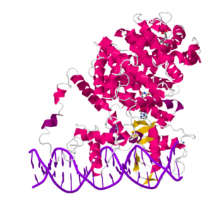
The modification of particular amino acids in nuclear receptors through phosphorylation, acetylation, ubiquitination, SUMOylation, and methylation has been confirmed. All of these regulate the activity of nuclear receptors and have implications both in the cellular biology and in the physiology of the organism.
An example would be the phosphorylation of the PPAR-gamma at serine 112. This phosphorylation diminishes the transcriptional activity of the PPAR-gamma and is apparently involved in insulin sensitivity.
[clear]
Phosphorylation
Phosphorylation is perhaps the most studied modification in these proteins. Most amino acids that can be phosphorylated are located in the A/B region, although we can also find some both in the DBD and in the LBD.
The effect of the phosphorylation will depend on the phosphorylated amino acid and on the affected receptor.
In some cases, phosphorylation increases transcriptional activity. In this sense, it has been proven that, in certain situations, the phosphorylation of the receptor can be responsible for its transcriptional activity, even in a ligand-independent manner.
On the other hand, some phosphorylations diminish transcriptional activity or can go as far as stopping it by separating the receptor and the DNA or the ligand.
Who phosphorylizes nuclear receptors?
Most phosphorylatable amino acids are serines surrounded by prolines. This sequence corresponds to the targets of proline-dependent kinases. Among them we find families as relevant as CDKs or MAPKs.
Images: Bromomir, Protein Data Bank
[one_half first]
[icon style=”default” size=”20″]fa-chevron-left[/icon] It Helps to Have Ligands in High Places
[/one_half]
[one_half]
Classification of Nuclear Receptors [icon style=”default” size=”20″]fa-chevron-right[/icon]
[/one_half]