Automatic Detection and Characterization of Atherosclerotic Plaque in Carotid Ultrasound
Cardiovascular diseases (CVD) are the leading cause of death in Western countries. The common basis of this group of diseases is atherosclerosis, a chronic degenerative process characterized morphologically by an asymmetric focal thickening of the innermost layer of the artery. The carotid arteries may develop atherosclerosis so that blood flow may be partially or totally blocked by the atherosclerotic plaque. The atherosclerotic plaque can be indirectly detected using carotid artery images. In this project, we have proposed a fully-automatic method to detect the presence of plaque in the far wall common carotid artery image [Zhang2015] and we are working in the extension of this method to other segments of the carotid.
Characterization of carotid plaque composition, more specifically the amount of lipid core, fibrous tissue, and calcified tissue, is an important task for the identification of plaques that are prone to rupture, and thus for early risk estimation of cardiovascular and cerebrovascular events. Due to its low costs and wide availability, carotid ultrasound has the potential to become the modality of choice for plaque characterization in clinical practice. However, its significant image noise, coupled with the small size of the plaques and their complex appearance, makes it difficult for automated techniques to discriminate between the different plaque constituents.

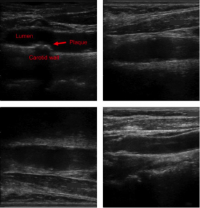
Figure: Four examples of carotid ultrasound images which contain plaques
In this project, we have addressed this challenging problem by exploiting the unique capabilities of the emerging deep learning framework. More specifically, and unlike existing works, which require a priori definition of specific imaging features or thresholding values, we propose to build a convolutional neural network (CNN) that will automatically extract from the images the information that is optimal for the identification of the different plaque constituents [Lekadir2016]. Based on our previous results, our goal is to progressively build a larger database of training cases together with our clinical partner to further enrich the plaque CNN classification as new datasets become available and to make it more robust to larger variability. We will also explore the relation of the plaque composition with the early prediction of cardiovascular and cerebrovascular events using other clinical datasets.
References:
[Zhang2015] Chen Zhang, Maria Del Mar Vila Muñoz, Petia Radeva, Roberto Elosua, María Grau, Angels Betriu, Elvira Fernandez-Giraldez and Laura Igual. Carotid Artery Segmentation in Ultrasound Images. CVII-STENT: Computing and Visualization for Intravascular Imaging and Computer Assisted Stenting in conjunction with MICCAI, 2015.
[Lekadir2016] Karim Lekadir, Alfiia Galimzianova, Àngels Betriu, Maria del Mar Vila, Laura Igual, Daniel L. Rubin, Elvira Fernández, Petia Radeva, and Sandy Napel. A Convolutional Neural Network for Automatic Characterization of Plaque Composition in Carotid Ultrasound. IEEE Journal of Biomedical and Health Informatics (J-BHI), 2016.
Last update: 25/11/2016